Vienna Surface Scanner Lab
Head: Assoc.Prof. Dr. Katrin Schäfer
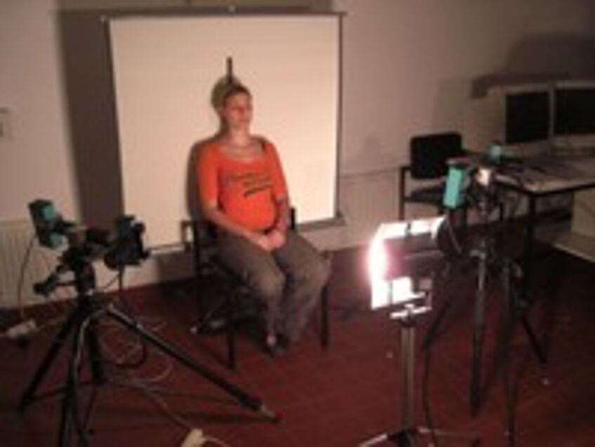
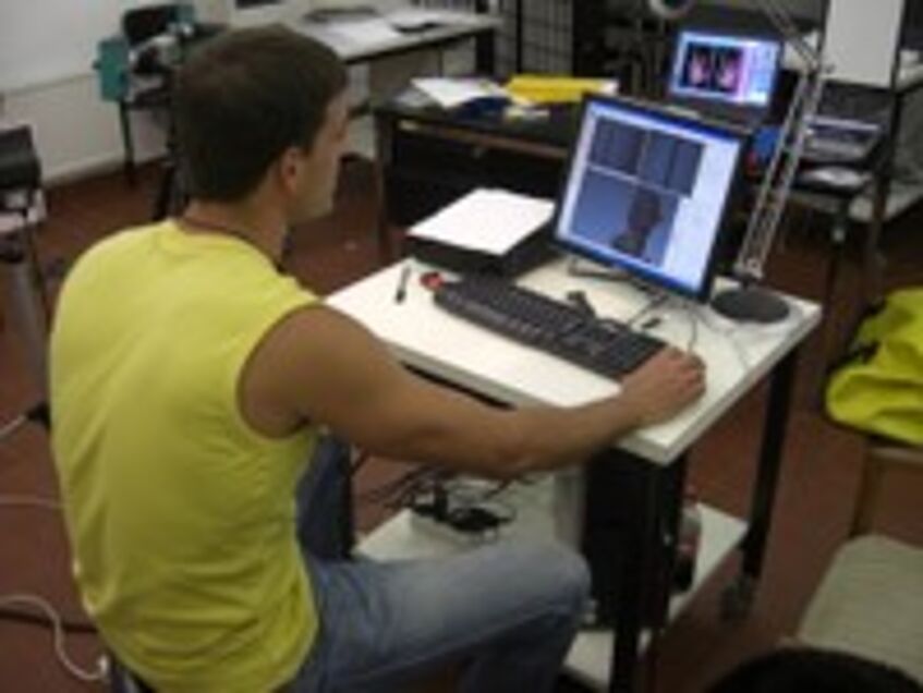
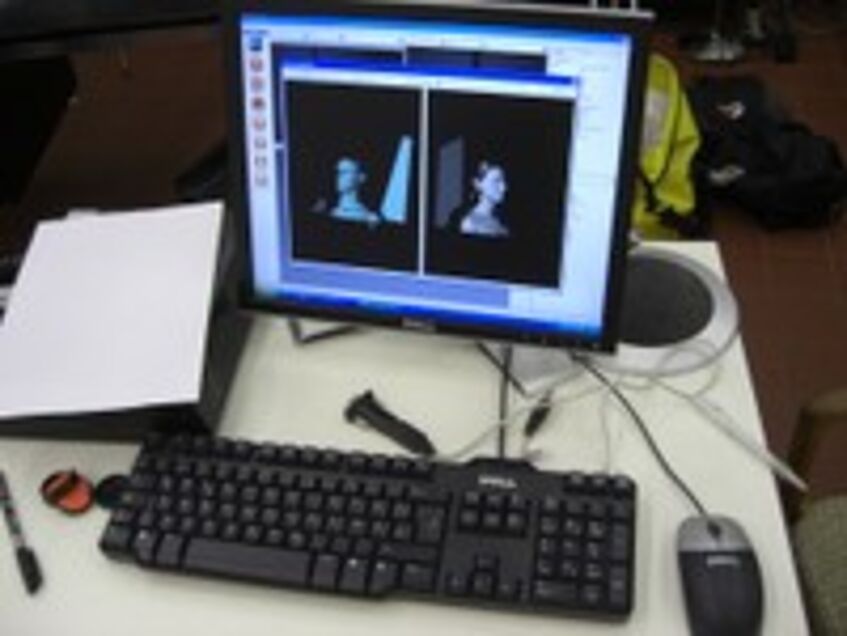
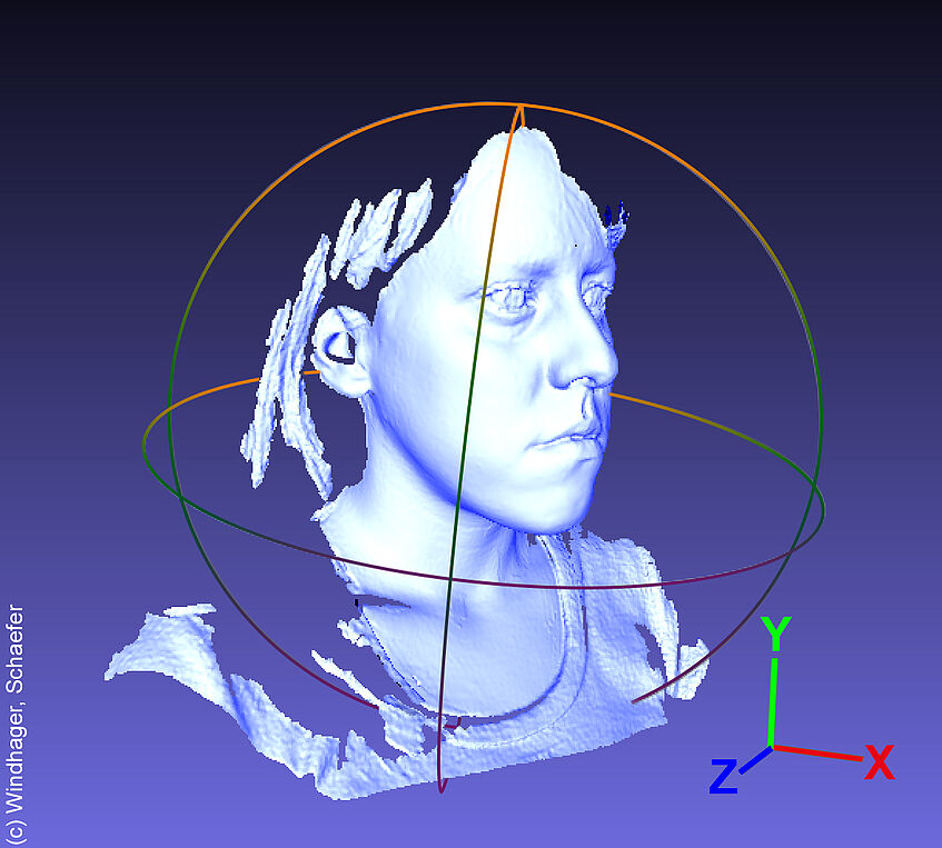
Surface scanning is a non-invasive method to capture the shape, texture and volume information of a three-dimensional object. In terms of accuracy, it can be as accurate as micro-tomography. Yet, it does not involve any exposure to ionizing radiation. Thus, this technology is highly suitable for sensitive material and living organisms. Both of our scanning systems – Breuckman OptoTop (www.aicon3d.de) and Artec (www.artec3d.com) 3D Scanners (MHT, Eva, Spider) – use active light projections. They provide color information and allow a high degree of mobility, including international travel. The main purposes of their use are archiving, presentation/visualization, and quantification. The latter is closely tied to the department’s strength in geometric morphometric methods. Furthermore, surface scans represent virtual specimens that can be easily shared, re-measured or addressed with new set of measurements. Also, surfaces can be averaged to depict a general trend in the data, or to reconstruct broken or otherwise incomplete specimens.
Recent equipment acquisition (granted by IP 547012 Morphogenetics in Comparative Anthropology to K. Schäfer and B. Wallner, 2015–2017, Univ. Vienna):
- Handheld 3D object scanner (Artec Eva)
- High resolution 3D scanner (Artec Spider)
Selected current and past projects:
- Face research (Schäfer Lab): Evolutionary theory explicitly places facial form in the middle of a causal chain as the mediating variable between biological causes of shape variation and person perception. Using geometric morphometrics, one can quantify and isolate those facial shape characteristics that lead to aesthetic and other judgments and directly compare them to the effects of physical processes (e.g., age, body height, BMI, hormones) on biological form ("Psychomorphospace", Schaefer et al. 2009).
Bravo Morante, G., Bookstein, F., Fischer, B., Schaefer, K., Aguilera, I. A., & Botella López, M. (2021).
Correlation of the human pubic symphysis surface with age-at-death: a novel quantitative method based on a bandpass filter.
International Journal of Legal Medicine, 135, 1935-1944.
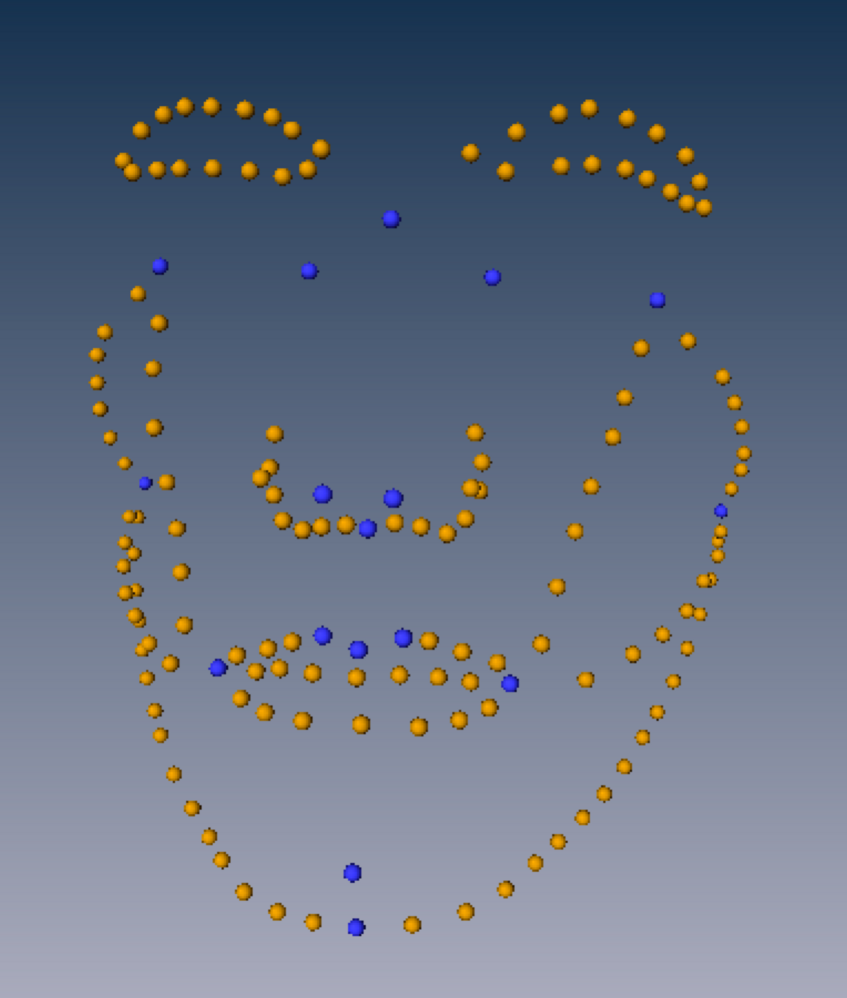
An example of geometric morphometric analyses: Fixed landmarks (blue) and sliding semilandmarks along curves (yellow) together with the whole mesh capture three-dimensional facial shape. After Procrustes superimposition all individual configurations are centered, scaled to the same size, and rotated to minimize the sum of squared distances over all landmarks. The resulting shape coordinates are then used in multivariate analyses such as shape regressions to relate facial shape to the predictor variable of interest such as a biological trait or a rating.
Reference: Schaefer K, Mitteroecker P, Fink B, Bookstein FL (2009). Psychomorphospace—from biology to perception, and back: Towards an integrated quantification of facial form variation. Biological Theory, 4(1), 98–106. rd.springer.com/article/10.1162/biot.2009.4.1.98
Modelling perineal swellings of female Barbary macaques
(Macaca sylvanus) (Sonnweber RS et al.)
In the course of this project, a new method was developed that enables the volumetric analysis of soft tissue when photogrammetric data is available. Artificial models of perineal swellings were used to evaluate the accuracy of this approach through the comparison of the photogrammetric data with surface scan data from the same models.
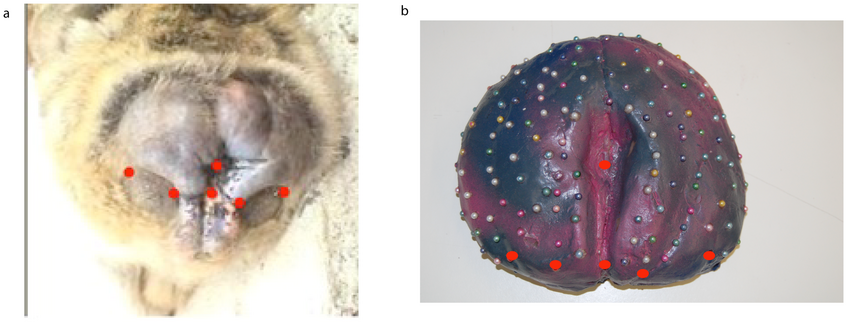
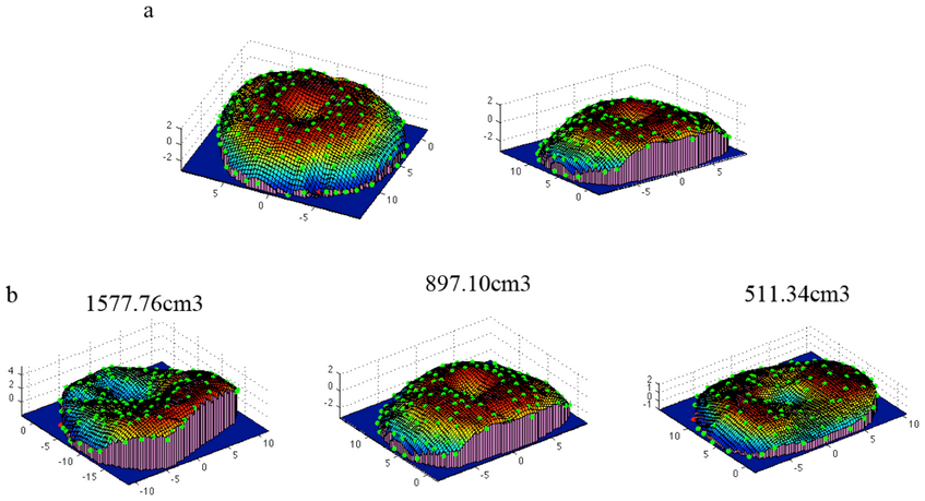
Reference and figure credit: Sonnweber RS, Stobbe N, Zavala Romero O, Slice DE, Fieder M, & Wallner B (2013) A new method for the analysis of soft tissues with data acquired under field conditions. PLoS ONE, 8(6), e67521. journals.plos.org/plosone/article
Quantification of entheses
(Pany D, Viola TB, Teschler-Nicola M)
More than twenty skeletons from Thunau/Gars am Kamp (Lower Austria, fortification with graveyard and settlement (early medieval)) that span a range of muscle mark development from faint to strong. The subsequent analyses focused on surface areas and perimeters, surface complexity measures, and planarity statistics.
Source including figures: https://www.uc.pt/en/cia/msm/Pany_Viola_Teschler.pdf
Variability of the femoral head in catarrhines – A new 3D method for describing anatomical structures
(Pfisterer T. et al.)
This project demonstrates that surface scans provide a valuable source of information in structures that lack multiple, reliable anatomic measurement points. For example, a sphere was fitted to the articular surface, and a cylinder to the shaft. Then the geometric properties of these simple geometric objects were used for multivariate analyses of species’ variation. [pdf]
Reference: Pfisterer T, Bookstein FL, Breuckmann B, Schaefer K, Viola TB, Woerner H, Seidler, H (2007) Variability of the femoral head in catarrhines – A new 3D method for describing anatomical structures. American Journal of Physical Anthropology, 44(Suppl.), 188–189
See also:
http://onlinelibrary.wiley.com/doi/10.1002/ajpa.22144/full
Sylvester AD & T Pfisterer (2012) Quantifying lateral femoral condyle ellipticalness in chimpanzees, gorillas, and humans. American Journal of Physical Anthropology, 149, 458–67. doi:10.1002/ajpa.22144.